Standing CT Scans Improve Diagnosis and Treatment of Lower-Extremity Injuries
Cesar de Cesar Netto, MD, Associate Professor of Orthopaedic Surgery, offers his expert opinion in a new DukeHealth article.
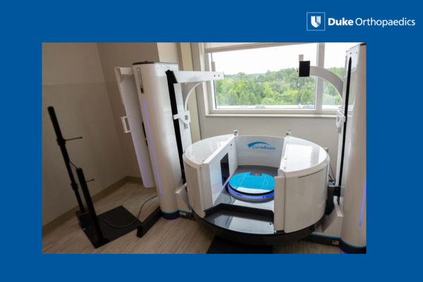
Duke Health is one of a few centers in North Carolina to offer a more accurate way to diagnose foot, ankle, knee, and hip problems with less radiation than traditional CT scans. Standing CT scans are taken while you are bearing weight and produce detailed 3D images that show how an injury affects alignment and functionality. This can help doctors decide whether surgery is needed, said Cesar de Cesar Netto, MD, a foot and ankle orthopaedic surgeon at Duke Health. “For example, with progressive collapsing flatfoot deformity, doctors can determine how far the deformity has progressed and if surgery or some other treatment is needed, and when,” he said. Read more.