About the Lab
The GRAV-ET Orthopaedics Lab, led by Dr. Cesar De Cesar Netto, MD, PhD, is dedicated to advancing foot and ankle care through cutting-edge translational and clinical research. Our work integrates state-of-the-art imaging, computational modeling, and advanced analytics to study lower extremity biomechanics during functional, weight-bearing activities. Using Weight-Bearing CT (WBCT) and 3D reconstructions, we accurately characterize alignment and deformity, complemented by tools such as plantar pressure mapping, dynamic gait analysis, and 3D printing for preoperative planning and patient-specific orthotics.
We also apply machine learning and predictive modeling to enhance diagnostic precision and identify markers of deformity progression. Clinically, our research spans retrospective and prospective studies on conditions such as Progressive Collapsing Foot Deformity (PCFD), multidirectional ankle instability, sports-related injuries, Hallux Valgus, midfoot and ankle arthritis, and other complex pathologies. By uncovering patient- and treatment-related factors that influence outcomes, we aim to inform evidence-based decisions and enable personalized care.
Our international, multidisciplinary team of surgeons, trainees, and scientists collaborates to bridge biomechanical modeling with clinical applications, delivering innovative solutions that elevate standards of care worldwide.
Research
Progressive Collapsing Foot Deformity (PCFD)
Progressive Collapsing Foot Deformity—formerly known as adult-acquired flatfoot or PTTD—is a complex, three-dimensional condition marked by collapse of the medial longitudinal arch, hindfoot valgus, forefoot abduction, and midfoot varus. This deformity results from failure of the posterior tibial tendon and key supporting ligaments, including the spring, deltoid, and talocalcaneal interosseous ligaments. PCFD affects approximately 5 million people in the U.S. and is staged based on flexibility and severity. Early stages may respond to orthoses, bracing, and physical therapy, while advanced cases often require surgical reconstruction or fusion. Weight-bearing CT and MRI are essential for accurate diagnosis and staging.
Multidirectional Ankle Instability
This condition involves symptomatic instability in two or more directions of ankle motion, often due to combined laxity of the medial and lateral ligaments and/or chronic syndesmotic injury. Unlike isolated lateral instability, surgical treatment typically addresses medial, lateral, and syndesmotic insufficiency. Patients commonly report persistent “giving way,” recurrent sprains, and functional limitations despite conservative care. Functional instability may also involve proprioceptive or neuromuscular deficits, even in the absence of mechanical laxity.
Hallux Valgus (Bunions)
Hallux valgus—commonly called a bunion—is a progressive deformity of the first ray, characterized by medial deviation of the first metatarsal and lateral rotation/pronation of the hallux. It presents as a painful prominence at the medial MTP joint and may be associated with hammertoes, calluses, and sesamoid shift. Risk factors include narrow or high-heeled footwear, genetics, abnormal biomechanics, and inflammatory conditions such as rheumatoid arthritis. Diagnosis relies on a clinical examination and weight-bearing radiographs to measure the hallux valgus angle (HVA) and intermetatarsal angle (IMA). Treatment ranges from footwear modifications and orthoses to surgical correction, tailored to the severity of the deformity.
Research Techniques
Weight-Bearing CT (WBCT)
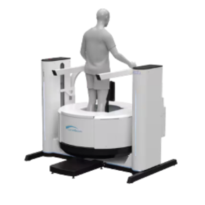
WBCT is a transformative imaging technology that captures accurate 3D measurements of the lower limb in a natural weight-bearing stance. At GRAV-ET, WBCT is central to our research, enabling precise assessment of alignment and deformity. Our lab utilizes the CurveBeamAI HiRise scanner, which provides comprehensive lower-limb imaging from toe tips to hips, supporting advanced modeling and surgical planning.
Advanced Visualization
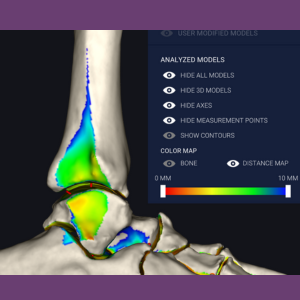
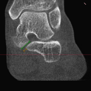
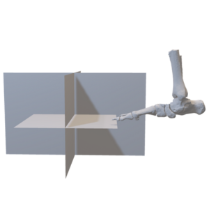
Advanced visualization enhances the diagnostic capabilities of WBCT combined with sophisticated 3D techniques in Computer Science to offer new insights into bone and joint physiology and pathology. Techniques such as Artificial Intelligence (AI) and Machine Learning (ML) are utilized. The Grav-ET lab is equipped with Disior software (https://www.disior.com/), which provides the research team with segmentation and advanced visualization tools, including distance measurement, coverage mapping, and automated 3D measurements. Hounsfield Unit (HU) analysis allows the study of local variations in Bone Mineral Density (BMD), named after Sir Godfrey Hounsfield, Nobel Prize winner for the invention of CT.
3D Printing
Our lab leverages 3D printing technology to create precise, patient-specific models of complex foot and ankle deformities. These models support preoperative planning, intraoperative guidance, and educational training, allowing surgeons to rehearse intricate procedures and improve surgical accuracy.
Surgical Planning
We provide clinicians and researchers with advanced visualization tools and 3D measurements to enable patient-specific surgical planning, ensuring tailored and effective treatment strategies.
Custom 3D-Printed Insoles
Custom orthotic insoles—developed through pressure mapping and 3D scanning—are designed to correct biomechanical abnormalities, redistribute plantar pressures, and alleviate pain in conditions such as plantar fasciitis and hallux valgus. Our lab uses technology from T-Soles, which uniquely integrates pressure scan data with WBCT imaging to create insoles customized to each patient’s bony anatomy. Research evaluates their effectiveness through quantitative gait and pressure analysis.
Pressure Scans
Pressure scanning provides detailed insights into plantar load distribution, identifying abnormal pressure zones and gait asymmetries. This technique facilitates the diagnosis of functional impairments, planning of surgical corrections, and evaluation of postoperative outcomes.
3D Gait Analysis
Three-dimensional gait analysis precisely measures foot and ankle motion during walking, revealing subtle biomechanical deviations linked to deformities or instability. It helps tailor treatment strategies and objectively evaluate surgical outcomes.
Anatomical Specimens Lab
Our cadaveric specimen lab enables biomechanical testing and anatomical validation of new surgical techniques and implant designs. By replicating clinical scenarios in a controlled setting, we generate critical evidence to support innovation in foot and ankle surgery.
People
Faculty Collaborators


Current Visiting Scholars


Resident Members
Medical Student Members


Past Members/Scholars


External Collaborators


Publications
Dr. Cesar Netto’s and GRAV-ET Labs' research publications leverage WBCT as a cornerstone for imaging, introducing novel 3D quantitative metrics (distance maps, coverage maps, and 3D angles) for analysis and integrating these with surgical decision-making. His methodologies range from high-tech imaging and computer modeling to cadaveric simulations, all aimed at a deeper understanding of flatfoot deformity and improving patient outcomes.
The Lab’s research approaches for multidirectional ankle instability combine cutting-edge imaging (WBCT with stress, 4DCT) to uncover hidden instabilities, computational analysis (distance mapping algorithms) to quantify them, and biomechanical experiments to validate these findings. His work emphasizes that not all ankle sprains are equal and that precise, multiplanar assessment is necessary to guide comprehensive treatment. By integrating these techniques, his contributions enable clinicians to identify which ankles require more than simple ligament repair and ensure that all aspects of instability are addressed to prevent long-term degeneration. In hallux valgus research, he merges high-fidelity imaging with practical surgical insights. By quantifying the deformity in three planes (and even at microscopic levels in cadavers), he has helped shift the paradigm of bunion assessment from simple angles on an X-ray to a comprehensive 3D evaluation. This, in turn, influences surgical planning and postoperative verification to improve long-term outcomes for patients with hallux valgus.
Wang CS, Huánuco Casas EJ, Luo EJ, Acker AS, Easley ME, de Cesar Netto C. A Comprehensive Weightbearing Computed Tomography Study on Patients With Hallux Valgus: Exploring Multiplanar Deformity Interrelationships. Foot Ankle Int. 2025 Mar;46(3):343-356. doi: 10.1177/10711007241309912. Epub 2025 Jan 24. PMID: 39861960.
Kruger KM, Lenz AL, Dibbern KN, de Cesar Netto C, Ledoux WR, Thorhauer ED, Burssens A, Siegler S, Rainbow MJ, Welte L, Peterson AC, Conconi M, Williams DE, Turmezei T, Hansen P, Lintz F, Leardini A. Standardizing 3 Dimensional Measurements in Foot and Ankle Imaging: A Contemporary Review and Methodological Proposal. Foot Ankle Clin. 2025 Mar;30(1):221-237. doi: 10.1016/j.fcl.2024.08.006. Epub 2024 Dec 3. PMID: 39894616; PMCID: PMC11788574.
Peterson AC, Requist MR, Benna JC, Nelson JR, Elhabian S, de Cesar Netto C, Beals TC, Lenz AL. Talar Morphology of Charcot-Marie-Tooth Patients With Cavovarus Feet. Foot Ankle Int. 2025 Mar;46(3):268-274. doi: 10.1177/10711007241309915. Epub 2025 Feb 12. PMID: 39937093.
Fayed AM, Jones M, de Carvalho KAM, Mansur NSB, de Cesar Netto C. Weightbearing Computed Tomography Measurements in Progressive Collapsing Foot Deformity: A Contemporary Review. Foot Ankle Orthop. 2025 Feb 26;10(1):24730114251316547. doi: 10.1177/24730114251316547. PMID: 40013107; PMCID: PMC11863214.
Grün W, Luo E, Pozzessere E, Huanuco Casas EJ, Acker A, Vermorel PH, Lintz F, de Cesar Netto C. Foot Malalignment and Proximal Fifth Metatarsal Fractures. Foot Ankle Int. 2025 May;46(5):561-570. doi: 10.1177/10711007251322141. Epub 2025 Mar 12. PMID: 40077926.
Anastasio AT, Wu KA, Luo EJ, Netto CC, Easley ME. Complications and Early-Term Radiographic Analysis of a Novel Active Compression Tibiotalocalcaneal Arthrodesis Nail With a Proximal Flexible Coil. Foot Ankle Orthop. 2025 Mar 18;10(1):24730114251323895. doi: 10.1177/24730114251323895. PMID: 40104093; PMCID: PMC11915544.
Lintz F, Pozzessere E, Grün W, Acker A, Huánuco Casas EJ, Ferkel E, de Cesar Netto C. A Hallux Valgus Surgical Planning Survey Using WBCT-based 3D Printing. Foot Ankle Orthop. 2025 Mar 18;10(1):24730114251325854. doi: 10.1177/24730114251325854. PMID: 40104096; PMCID: PMC11915313.
Talaski GM, Mallavarapu V, Behrens A, Mansur N, Aiyer A, Anastasio AT, Lintz F, de Cesar Netto C. Semi-automated Segmentation and Distance Mapping of Weight Bearing CT Data to Estimate Select Midfoot Joint Volumes. Foot Ankle Int. 2025 Jun;46(6):644-651. doi: 10.1177/10711007251328693. Epub 2025 Apr 22. PMID: 40261007.
Godoy-Santos AL, Teixeira Lobo CF, Rosemberg DL, Sposeto RB, Lintz F, de Cesar Netto C. Subtle tarsometatarsal ligament injury in a professional ballerina: weight-bearing computed tomography technical tip. Journal of the Foot & Ankle. 19, 1 (Apr. 2025), 1–7. DOI:https://doi.org/10.30795/jfootankle.2025.v19.1868.
Forin Valvecchi T, Marcolli D, De Cesar Netto C. Advanced Three-Dimensional Assessment and Planning for Hallux Valgus. Foot Ankle Clin. 2025 Jun;30(2):349-362. doi: 10.1016/j.fcl.2024.06.008. Epub 2024 Dec 30. PMID: 40348467.
Lam P, Fletcher L, Watt C, Ray R, Dalmau-Pastor M, de Cesar Netto C, Lewis TL. First Metatarsal Pronation Correction After Fourth-Generation Percutaneous Transverse Osteotomy for Hallux Valgus. Foot Ankle Int. 2025 Jun 28:10711007251344273. Doi: 10.1177/10711007251344273. Epub ahead of print. PMID: 40580076.
Mansur NSB, Hume D, Kwon J, de Carvalho KAM, Dibbern K, de Cesar Netto C. Correction of syndesmotic malreduction following fixation flexibilization. Sci Rep. 2025 Jul 1;15(1):22434. doi: 10.1038/s41598-025-04117-x. PMID: 40593952; PMCID: PMC12218112.
Herrera-Pérez M, Valderrabano V, Netto CC, I Ferkel E, Leme-Godoy A, Dway A, Pais-Brito JL, Tejero S. Mid-to-Long-term Clinical Outcomes of Ankle Arthroscopy on the Treatment of Chronic Ankle Conditions: Systematic Review and Meta-analysis. Foot Ankle Spec. 2025 Jul 2:19386400251345536. Doi: 10.1177/19386400251345536. Epub ahead of print. PMID: 40598973.
Lintz F, Pozzessere E, Carfì C, Riewruja K, Grün W, Cornelius C, Ellis S, Netto CC. Reconstruction of the Interosseous Talocalcaneal Ligament in Progressive Collapsing Foot Deformity: A Review of the Literature and Description of a Novel Surgical Technique. J Foot Ankle Surg. 2025 Jul 7:S1067-2516(25)00193-0. doi: 10.1053/j.jfas.2025.06.008. Epub ahead of print. PMID: 40633712.
Lintz F, Grün W, Pozzessere E, Luo E, Huanuco Casas EJ, Vermorel PH, Acker A, de Cesar Netto C. Fifth Metatarsal Stress Fractures Are Associated With Increased Bone Density and Altered Alignment on Weight-bearing CT. Clin Orthop Relat Res. 2025 Jul 10. Doi: 10.1097/CORR.0000000000003613. Epub ahead of print. PMID: 40658443.
Zambelli R, Santos AF, Moreira LR, Ribeiro HM, Simões R, Magalhães JM, Constantino P, Salomão MC, Netto CC, Leopoldino AO. Bacterial profile and antimicrobial resistance in diabetic foot ulcer infections: a 10-year retrospective cohort study. Braz J Infect Dis. 2025 Jul 31;29(5):104570. doi: 10.1016/j.bjid.2025.104570. Epub ahead of print. PMID: 40749580.
De Cesar Netto C, Barbachan Mansur NS, Talaski G, Jasper RP, Schmidt E, Mendes de Carvalho KA, Dibbern K, Lintz F, Ellis SJ, Anderson DD. From Asymptomatic Flatfoot to Progressive Collapsing Foot Deformity: Peritalar Subluxation Is the Main Driver of Symptoms. J Bone Joint Surg Am. 2025 Aug 4. doi: 10.2106/JBJS.24.01619. Epub ahead of print. PMID: 40758778Lalevéeée M, Khvesyuk M, Behrens A, Beaudet P, Perrier A, DaCosta A, Lintz F, Dibbern K, de Cesar Netto C. Valgus Deviation of the Intersesamoid Crista in Hallux Valgus and Its Association With the Distal Metatarsal Articular Angle: A Pilot Study. Foot Ankle Orthop. 2025 Aug 16;10(3):24730114251355501. doi: 10.1177/24730114251355501. PMID: 40827130; PMCID: PMC12357992.
Bernasconi ALalevéeée M, Fernando C, Izzo A, de Cesar Netto C, Lintz F. Early Tibiotalar Joint Changes Following Subtalar Arthrodesis for Progressive Collapsing Foot Deformity: A 3D Distance Mapping Short Scientific Report. Foot Ankle Int. 2025 Sep 1:10711007251361123. Doi: 10.1177/10711007251361123. Epub ahead of print. PMID: 40888339Lalevéeée M, Dagneaux L, Lintz F, de Cesar Netto C. Flatfoot: New diagnostic modalities. Orthop Traumatol Surg Res. 2025 Sep 9:104415. Doi: 10.1016/j.otsr.2025.104415. Epub ahead of print. PMID: 40935328.
Mansur NSB, Fayed A, Chinelati R, Schmidt ELalevéeee M, de Cesar Netto C. Allograft Bone-Block Plantarflexion First Tarsometatarsal Arthrodesis: Short-term Outcomes. Foot Ankle Int. 2025 Sep 12:10711007251363926. Doi: 10.1177/10711007251363926. Epub ahead of print. PMID: 40938130.
Mallavarapu V, Carvalho KAM, Behrens A, Jones MT, Jasper R, Zeller H, Dibbern K, de Cesar Netto C. Weightbearing CT 3-D Mapping of First Metatarsophalangeal and Sesamoid Joint Interactions in Hallux Valgus: A Case-Control Study. Foot Ankle Int. 2025 Sep 27:10711007251363257. Doi: 10.1177/10711007251363257. Epub ahead of print. PMID: 41014167.
Grün W, Pozzessere E, Luo EJ, Mansur NSB, Acker A, Vermorel PH, Lintz F, Netto CC. More than meets the eye? Evaluating 3D printing for progressive collapsing foot deformity classification. J Foot Ankle Surg. 2025 Sep 25; S1067-2516(25)00283-2. doi: 10.1053/j.jfas.2025.09.004. Epub ahead of print. PMID: 41015331.
Grün W, Vermorel PH, Luo EJ, Yang D, Pozzessere E, Talaski GM, Lintz F, de Cesar Netto C. Three-Plane Alignment of the Second Metatarsal Improves Reliability of Weightbearing CT Measurements in Lisfranc Injury Assessment. Foot Ankle Orthop. 2025 Sep 25;10(3):24730114251372593. doi: 10.1177/24730114251372593. PMID: 41018049; PMCID: PMC12464389.
Correction of syndesmotic malreduction following fixation and flexibilization. Mansur NSB, Hume D, Kwon J, de Carvalho KAM, Dibbern K, de Cesar Netto C.Sci Rep. 2025 Jul 1;15(1):22434. doi: 10.1038/s41598-025-04117-x.PMID: 40593952
Advanced Three-Dimensional Assessment and Planning for Hallux Valgus. Forin Valvecchi T, Marcolli D, De Cesar Netto C.Foot Ankle Clin. 2025 Jun;30(2):349-362. doi: 10.1016/j.fcl.2024.06.008. Epub 2024 Dec 30.PMID: 40348467
Semi-automated Segmentation and Distance Mapping of Weight-Bearing CT Data to Estimate Select Midfoot Joint Volumes.Talaski GM, Mallavarapu V, Behrens A, Mansur N, Aiyer A, Anastasio AT, Lintz F, de Cesar Netto C.Foot Ankle Int. 2025 Jun;46(6):644-651. doi: 10.1177/10711007251328693. Epub 2025 Apr 22.PMID: 40261007
Standardizing 3 Dimensional Measurements in Foot and Ankle Imaging: A Contemporary Review and Methodological Proposal. Kruger KM, Lenz AL, Dibbern KN, de Cesar Netto C, Ledoux WR, Thorhauer ED, Burssens A, Siegler S, Rainbow MJ, Welte L, Peterson AC, Conconi M, Williams DE, Turmezei T, Hansen P, Lintz F, Leardini A.Foot Ankle Clin. 2025 Mar;30(1):221-237. doi: 10.1016/j.fcl.2024.08.006. Epub 2024 Dec 3. PMID: 39894616
Cadaveric Diagnostic Study of Subtle Syndesmotic Instability Using a 3-Dimensional Weight-Bearing CT Distance Mapping Algorithm. de Cesar Netto C, Barbachan Mansur NS, Talaski G, Behrens A, Mendes de Carvalho KA, Dibbern K.J. Bone Joint Surg Am. 2025 Feb 19;107(4):397-407. doi: 10.2106/JBJS.24.00199. Epub 2024 Dec 19.PMID: 39700305
Anatomical and Micro-CT Assessment of the First Metatarsal Head Vascularization and Soft Tissue Envelope Following Minimally Invasive Chevron Osteotomy for Hallux Valgus Deformity. Carvalho KAM, Fayed A, Barbachan Mansur NS, Godoy-Santos AL, Talusan P, Chrea B, de Cesar Netto C, Johnson AH, Dalmau-Pastor M.Foot Ankle Int. 2025 Jan;46(1):102-114. doi: 10.1177/10711007241298681. Epub 2024 Nov 29.PMID: 39611439
Ankle osteoarthritis: Toward new understanding and opportunities for prevention and intervention. Anderson DD, Ledoux WR, Lenz AL, Wilken J, Easley ME, de Cesar Netto C.J Orthop Res. 2024 Dec;42(12):2613-2622. doi: 10.1002/jor.25973. Epub 2024 Sep 13.PMID: 39269016
Hindfoot Alignment in Flexible Cavovarus Deformity Under Orthostatic and Coleman Block Test Positions: A Weight-bearing Computed Tomography Study. Pires EA, Lobo CFT, Fonseca FC, Sposeto RB, Barbachan Mansur NS, Easley ME, de Cesar Netto C, Godoy-Santos AL.Foot Ankle Int. 2024 Sep;45(9):1027-1037. doi: 10.1177/10711007241258180. Epub 2024 Jul 26.PMID: 39056577
Deformities Influencing Different Classes in Progressive Collapsing Foot. Fayed A, Mallavarapu V, Schmidt E, de Carvalho K, Lalevé M, Kim KC, Ehret A, Rojas EO, Lintz F, Ellis SJ, Mansur NS, de Cesar Netto C.Iowa Orthop J. 2023 Dec;43(2):8-13.PMID: 38213846
Research Opportunities
If you have any questions about our research, please let us know! If you're interested in any of our research opportunities, send your CV, a cover letter describing your research interests and experiences, and contact information for two references.
International Fellowship Program
- Lead independent research on complex foot and ankle deformities.
- Shadow Dr. Netto and Duke Foot and Ankle Faculty in the clinic and operating room.
- Collaborate with a multidisciplinary team of surgeons, researchers, and trainees.
- Mentor trainees, graduate, and undergraduate students through the research process.
Graduate and Undergraduate Research Positions
- Gain hands-on experience in foot and ankle research projects with expert mentors.
- Study complex foot and ankle deformities.
- Learn advanced skills in imaging analysis, interpretation, 3D modeling, and data science.
- Contribute meaningfully to translational and clinical research projects.
Visiting Scholars Program
- Participate in short-term research stays, share knowledge and techniques.
- Collaborate on ongoing projects and introduce new perspectives.
- Shadow Dr. Netto and Duke Foot and Ankle Faculty in the clinic and operating room.
- Present your research and engage in lab meetings.
Collaborative Research Projects
- Work on multidisciplinary research initiatives.
- Share expertise in foot and ankle and imaging-based technologies to advance innovative research.
- Participate in joint grant applications and publications.
News & Events
- WBCT Society
- Past Events
- Webinars